Scientific Instrument
CHEETAH™ : Light Field Microscopic Add-on Module
For demo, presentation and customized application, please contact us
Contact to the team
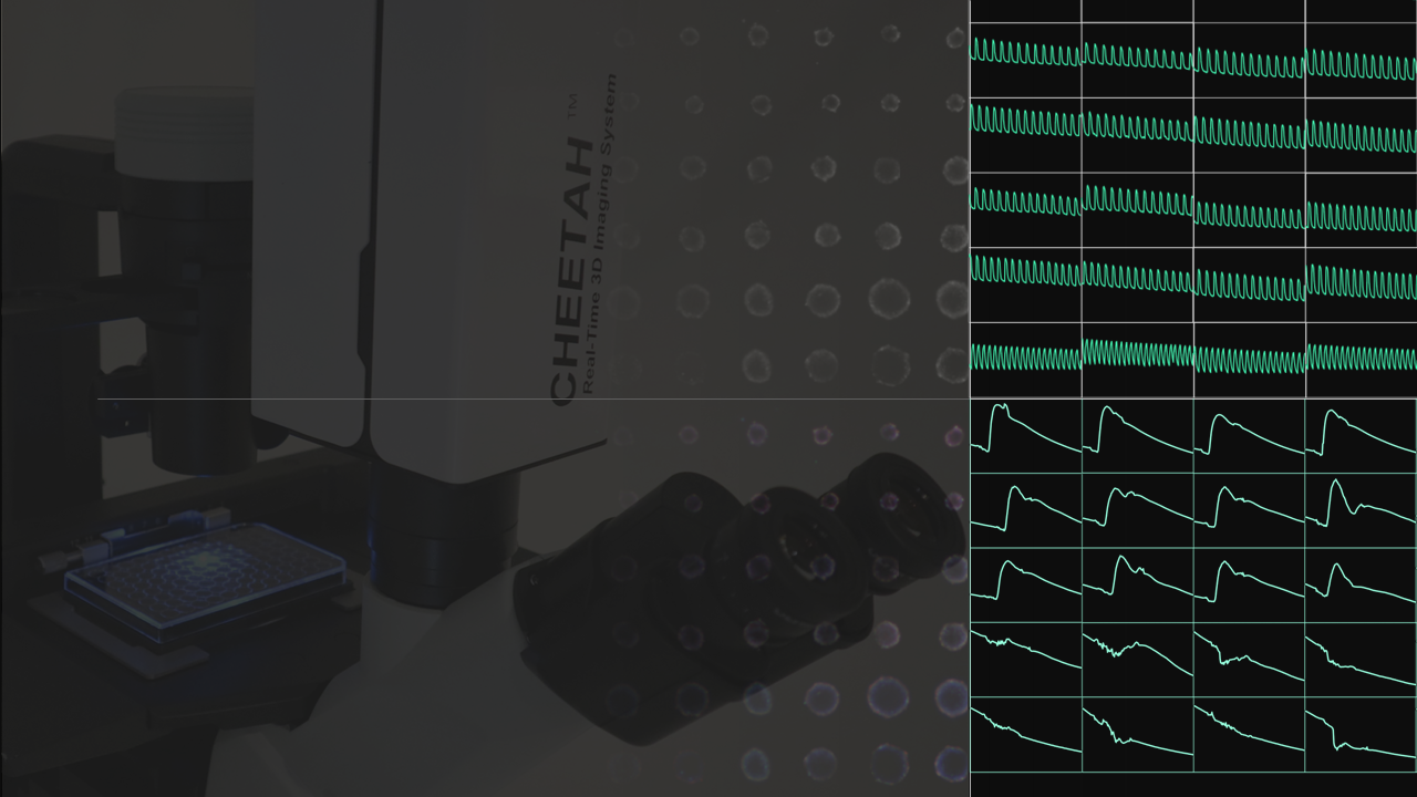
Detecting The Undetectable In Real-time 3D
Analyze mechanism-of-action (MOA) in depth of organotypic 3D cell culture with unprecedented volumetric frame rate (>30fps)
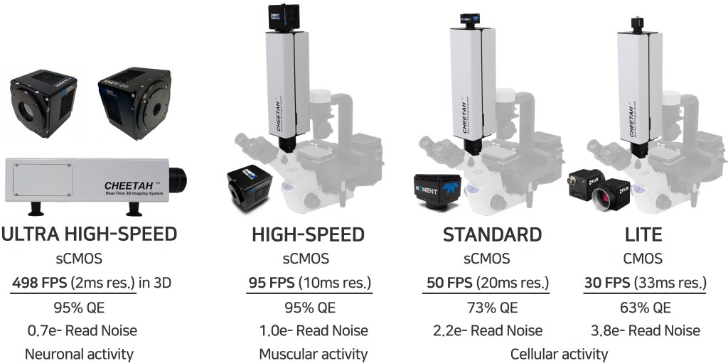
- Neuronal activity
- Cardiac/Muscular activity
- Immune activity
- Complex functional activity
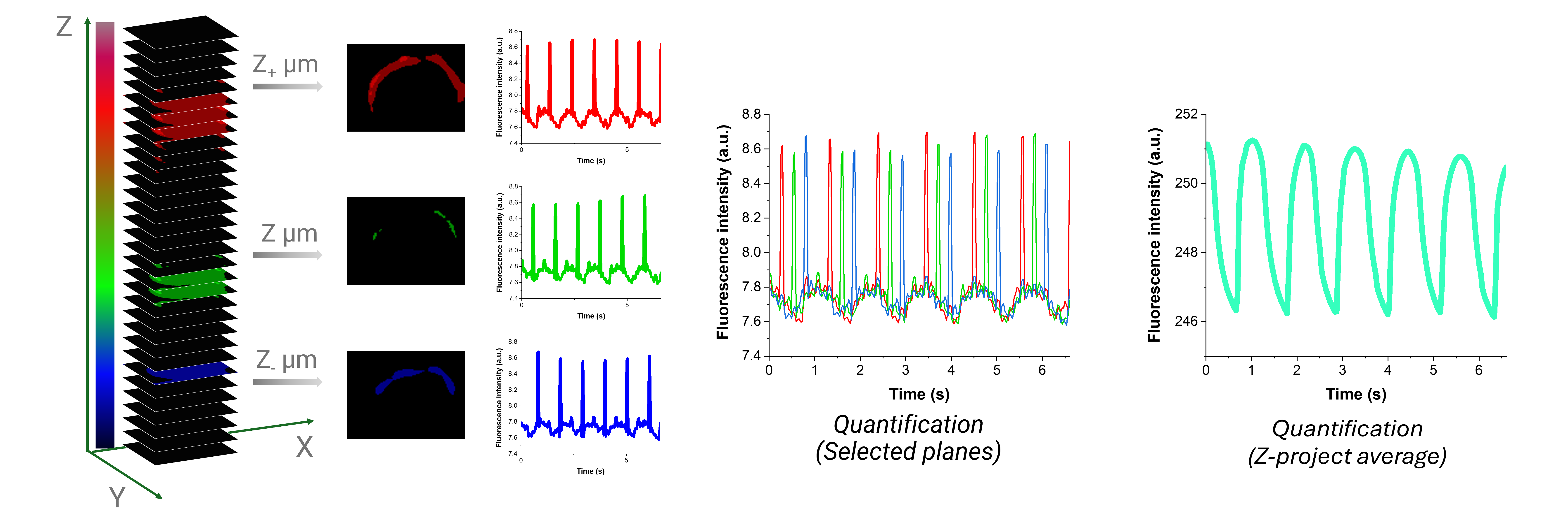
- Extractability of a single-shot 3D image in 32 stacks each frame and individual intensity data point
Scientific Camera Selection At Your Needs
Seamless Installation On Wide-field Fluorescent Microscope

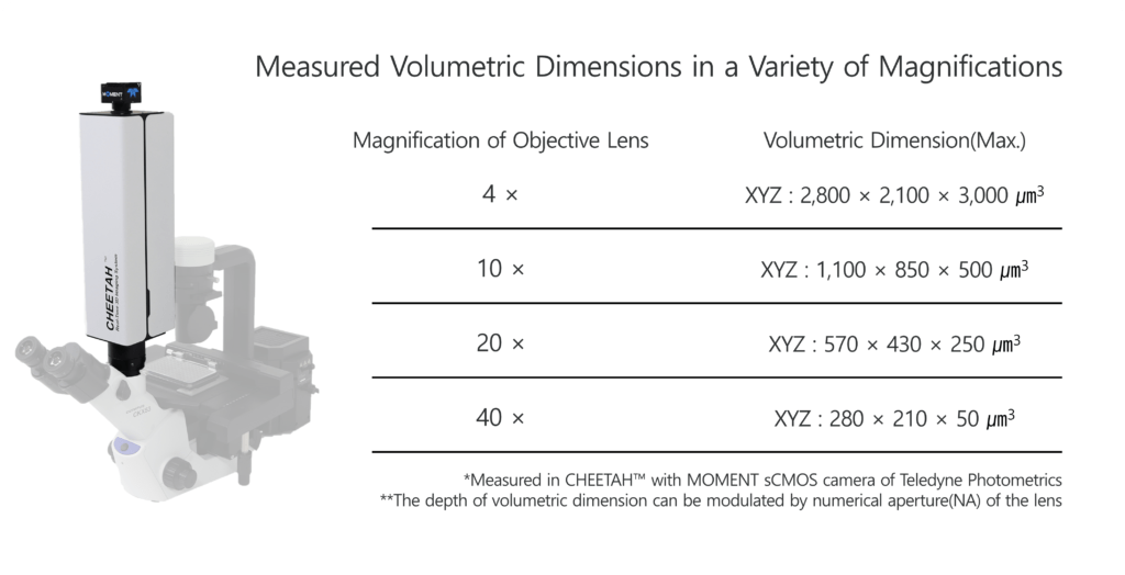
Volumetric Dimensions For Your Interest
The System was Optimized in
CPU : AMD Ryzen Threadripper PRO 3955WX
GPU : NVIDIA RTX A5000 D6
RAM : 128 GB
CHEETAH™ : Applications
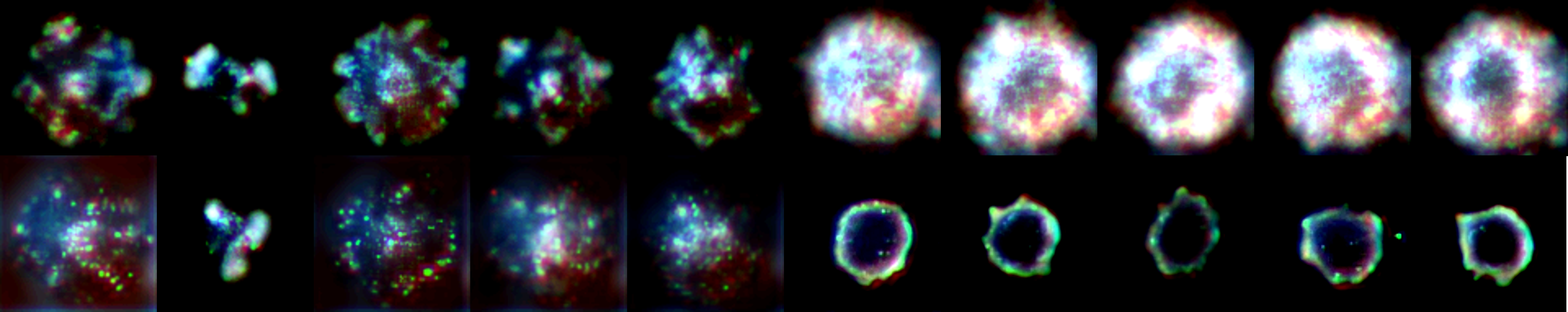
Calcium imaging of Fluo-4 Labeled HEK293 3D spheroid
Human embryonic kidney (HEK293) cells are widely used for the heterologous expression of voltage- and ligand-gated ion channels. Fluo-4, calcium ion indicator, was labeled to visualize and monitor changes in intracellular calcium levels. This video includes in observation the calcium dynamics of HEK293 spheroid in 10 fps 3D.
Dose response GPCR assay on HEK293 spheroids
- Real-time 3D observations (10fps in 3D) and measurements to analyze how calcium indicator labeled HEK293 spheroids responding to varying doses of carbachol, an agonist that binds to muscarinic acetylcholine receptors (mAChRs), a subfamily of G-protein coupled receptors (GPCR) expressed on the cells.
- Calcium signaling in GPCR is a critical process involving increasing the concentration of calcium ions in the cytoplasm to initiate cellular events.
Cardiotoxicity assay on iPSC-derived cardiomyocyte 3D spheroids
- Real-time 3D observations and measurements of cardiac 3D spheroids (Only 20K cells per well) for cardiotoxicity assay, utilizing NEXEL’s Cardiosight®-S in calcium imaging and voltage imaging with an interval time of 33ms and 11ms respectively.
- By combining calcium imaging’s prowess in tracking cellular activity with voltage imaging’s ability to monitor membrane potential, we gained unprecedented insights into the electrophysiological behavior of 3D cardiac miniatures with different doses of drug, potassium channels blocker, E-4031.
